The structures of biotin, biocytin, and carboxybiotin (the active metabolic intermediate) are shown in Figure 13. Biotin is widely distributed in many foods as biocytin (ε-amino-biotinyllysine), which is released on proteolysis. It is synthesized by intestinal flora in excess of requirements. Deficiency is unknown, except among people maintained for many months on total parenteral nutrition, and a very small number who eat abnormally large amounts of uncooked egg white, which contains avidin, a protein that binds biotin and renders it unavailable for absorption (Murray et. al. 2009).
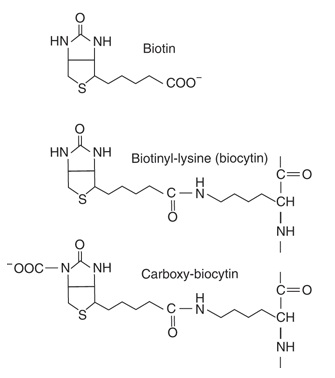
Figure 13. Biotin, biocytin, and carboxy-biocytin. Harper’s Illustrated Biochemistry 28 ed. (p. 953), by R. K. Murray, D. A. Bender, K. M. Botham, P. J. Kennelly, V. W. Rodwell & P. A. Well, China: McGraw-Hill Companies. Copyright 2009 by McGraw-Hill Companies.
|
Biotin (Figure 14) functions to transfer carbon dioxide in a small number of reactions: acetyl-CoA carboxylase, pyruvate carboxylase , propionyl-CoA carboxylase, and methylcrotonyl-CoA carboxylase. A holocarboxylase synthetase catalyzes the transfer of biotin onto a lysine residue of the apo-enzyme to form the biocytin residue of the holoenzyme. The reactive intermediate is 1-N -carboxy-biocytin, formed from bicarbonate in an ATP-dependent reaction. The carboxy group is then transferred to the substrate for carboxylation. Biotin also has a role in regulation of the cell cycle, acting to biotinylate key nuclear proteins (Murray et. al. 2009).
|
|
According to Murray et. al. (2009), biotin deficiency does not occur naturally because the vitamin is widely distributed in food. Also, a large percentage of the biotin requirement in humans is supplied by intestinal bacteria. However, the addition of raw egg white to the diet as a source of protein induces symptoms of biotin deficiency, namely, dermatitis, glossitis, loss of appetite, and nausea. Raw egg white contains a glyco protein, avidin, which tightly binds biotin and prevents its absorption from the intestine. With a normal diet, however, it has been estimated that 20 eggs/day would be required to induce a deficiency syndrome. Thus, inclusion of an occasional raw egg in the diet does not lead to biotin deficiency, although eating raw eggs is generally not recommended due to the possibility of salmonella infection (Harvey & Ferrier 2011).
|
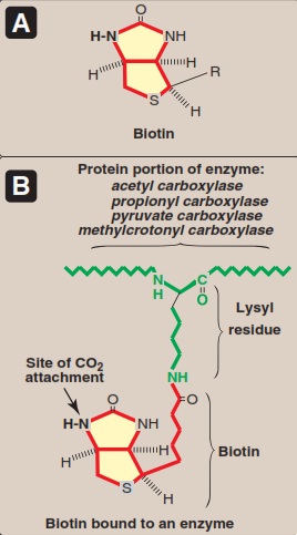
Figure 14. A. Structure of biotin. B. Biotin covalently bound to a lysyl residue of a biotin-dependent enzyme. Lippincott’s Illustrated Reviews: Biochemistry 5th ed. (p. 381), by R. A. Harvey & D. R. Ferrier, 2011, Philadelphia: Lippincott Williams & Wilkins, a Wolters Kluwer business. Copyright 201 by Lippincott Williams & Wilkins, a Wolters Kluwer business.
|
